Herpes Simplex Virus is a common pathogen and its primary infection is usually asymptomatic. There
are two immunologically distinct types of HSV: Type 1 and Type 2. HSV 1 is generally associated with
oral infection and lesions above the waist, and HSV 2 is associated with genital infections and lesions
below the waist. Clinical cases primarily are 1) eczema herpeticum with eczematous skin changes with
numerous lesions, 2) Gingivo-stomatitis and 3) Herpes sepsis, almost only found in newly born of premature infants. DIAGNOSTIC AUTOMATION ELISA HSV IgM is an accurate serologic method to
detect HSV specific antibody IgM in serum sample.
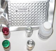
Purified HSV antigen is coated on the surface of microwells. Diluted patient serum is added to wells,
and the HSV IgM specific antibody, if present, binds to the antigen. All unbound materials are washed
away. After adding enzyme conjugate, it binds to the antibody-antigen complex. Excess enzyme conjugate is washed off and TMB Chromogenic substrate is added. The enzyme conjugate catalytic
reaction is stopped at a specific time. The intensity of the color generated is proportional to the amount
of IgM specific antibody in the sample. The results are read by a microwell reader compared in a parallel manner with calibrator and controls.
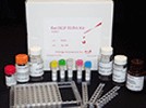
HSV-1 2 IgM ELISA Kit is an enzyme linked immunosorbent assay (ELISA) for the diction of IgM class antibodies to HSV-1 in human serum or plasma. HSV-1 and 2 are virtually identical, sharing approximately 50% of their DNA and have over 80% of common antigens. Both types infect the body's mucosal surfaces, usually the mouth or genitals, and then establish latency in the nervous system. For both types, at least two-thirds of infected people have no symptoms, or symptoms too mild to notice. However, both types can recur and spread even when no symptoms are present. By the time they're teenagers or young adults, about 50% of Americans have HSV-1 antibodies in their blood. By the time they are over age 50, some 80-90% of Americans has HSV-1 antibodies. By comparison, almost all HSV-2 is encountered after childhood, when people become sexually active. HSV type 1 is the cause of most orofacial herpes and HSV encephalitis; type 2 is the primary cause of initial and recurrent genital herpes and neonatal HSV. Reactivation of latent HSV infection is a frequent complication of immunosuppression due to cancer, transplantation and AIDS. Asymptomatic genital shedding of HSV-2 is more common than HSV-1 and occurs more frequently during the first 3 months after acquisition of primary type 2 disease than during later periods. The presence of HSV IgG antibody is indicative of previous exposure A significant increases in HSV IgG is an indicative of reactivation, current or recent infection. IgM antibody is present after primary HSV infection.
More about: HSV-1 2 IgM ELISA Kit sale
Read more: Reagent Product